the Preimplantation period
Stage 1 of Human Development
{WEEK 1}
Read:
Note: You can follow along in your Histology and Embryology textbook if you want to, beginning on page 19 with Chapter 3.
Prenatal development starts at the beginning of pregnancy or from the time that conception takes place. At the time of conception, fertilization of the egg, which is the female sex cell, occurs. The egg is penetrated by a single sperm, which is the male sex cell. The result is the formation of a zygote or fertilized egg.
Watch:
Below is a YouTube video that shows the process of fertilization, the beginning of life!
Video: Nucleus Medical Media. "Fertilization." Online video clip. YouTube. YouTube, 31 Jan. 2013. Web 24 Sept. 2013.
Read:
During fertilization, as you just saw, meiosis occurs within the zygote. Meiosis involves the process of joining the genetic information or chromosomes of both the egg and sperm. The result is a new individual with "shuffled" chromosomes, gaining half of it's genetic make-up from each parent.
After meiosis is complete, the zygote then begins to grow undergoing mitosis or cleavage, which is individual cell division. What started out as 1 zygote soon becomes 2 cells, then 4, then 8, and so on. When it reaches a size of about 16 cells that are tightly packed together, the zygote then is termed a morula. The morula continues dividing and in the process the cells secrete some fluid. Now the morula resembles a vesicle that is referred to as a blastocyst.
As the blastocyst continues to grow and divide (undergoing mitosis), it travels from the site where fertilization took place (usually the fallopian tube) to the uterus. By the end of the first week, the blastocyst stops traveling and undergoes implantation and thus becomes embedded in the uterine wall. The implanted blastocyst will now consist of an outer later of cells referred to as the trophoblast layer, which will develop into the placenta and other prenatal support tissue, and an inner mass of cells referred to as the embryoblast layer, which will develop into the embryo in the next period of development.
Look:
Below is a picture where you can see the zygote changing and moving to the uterus where it implants. Notice the trophoblast and embryoblast layers which are evident after implantation has occurred.
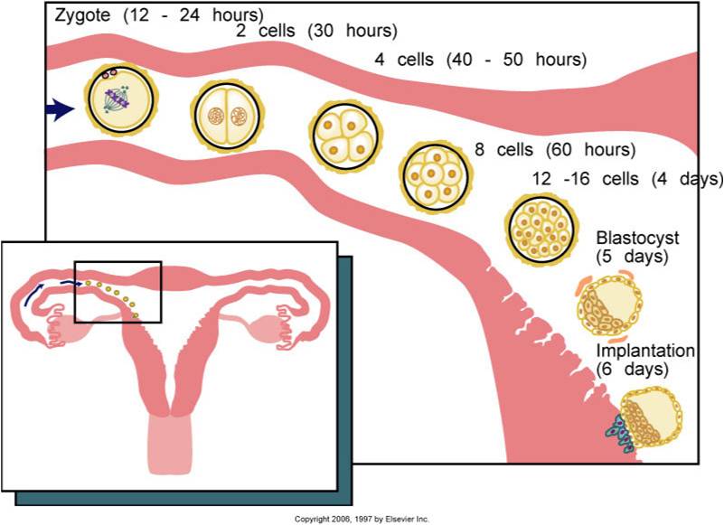
Image: Bath-Balough, M. and Fehrenbach, M. J. Illustrated Dental Embryology, Histology, and Anatomy. 2011. Diagram. Figure 3-3. 22.
Activities:
Now it's time for you to practice and see what you have learned. Please continue on to the Activity Page using the "Next Page" button. To go back to the previous page use the "Back" button.
Activities:
Now it's time for you to practice and see what you have learned. Please continue on to the Activity Page using the "Next Page" button. To go back to the previous page use the "Back" button.
